Selected Publications

Independent compartmentalization of functional, metabolic, and transcriptional maturation of hiPSC-derived cardiomyocytes
Human induced pluripotent stem cell-derived cardiomyocytes (hiPSC-CMs) recapitulate numerous disease and drug response phenotypes, but cell immaturity may limit their accuracy and fidelity as a model system. Cell culture medium modification is a common method for enhancing maturation, yet prior studies have used complex media with little understanding of individual component contribution, which may compromise long-term hiPSC-CM viability. Here, we developed high-throughput methods to measure hiPSC-CM maturation, determined factors that enhanced viability, and then systematically assessed the contribution of individual maturation medium components. We developed a medium that is compatible with extended culture. We discovered that hiPSC-CM maturation can be sub-specified into electrophysiological/EC coupling, metabolism, and gene expression and that induction of these attributes is largely independent. In this work, we establish a defined baseline for future studies of cardiomyocyte maturation. Furthermore, we provide a selection of medium formulae, optimized for distinct applications and priorities, that promote measurable attributes of maturation.
Cell Rep,
2024
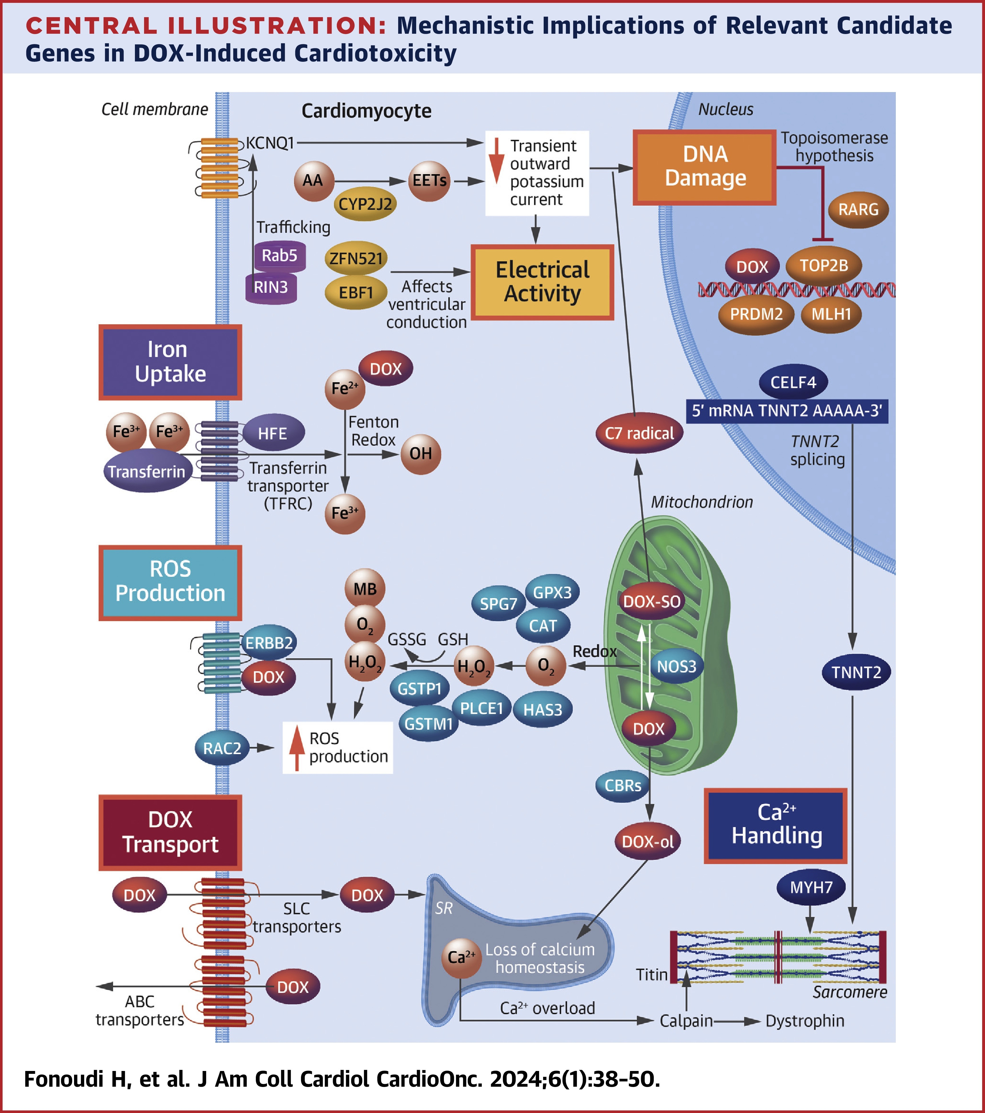
Functional validation of doxorubicin-induced cardiotoxicity-related genes
Background: Genome-wide association studies and candidate gene association studies have identified more than 180 genetic variants statistically associated with anthracycline-induced cardiotoxicity (AIC). However, the lack of functional validation has hindered the clinical translation of these findings. Objectives: The aim of this study was to functionally validate all genes associated with AIC using human induced pluripotent stem cell-derived cardiomyocytes (hiPSC-CMs). Methods: Through a systemic literature search, 80 genes containing variants significantly associated with AIC were identified. Additionally, 3 more genes with potential roles in AIC (GSTM1, CBR1, and ERBB2) were included. Of these, 38 genes exhibited expression in human fetal heart, adult heart, and hiPSC-CMs. Using clustered regularly interspaced short palindromic repeats/Cas9-based genome editing, each of these 38 genes was systematically knocked out in control hiPSC-CMs, and the resulting doxorubicin-induced cardiotoxicity (DIC) phenotype was assessed using hiPSC-CMs. Subsequently, functional assays were conducted for each gene knockout on the basis of hypothesized mechanistic implications in DIC. Results: Knockout of 26 genes increased the susceptibility of hiPSC-CMs to DIC. Notable genes included efflux transporters (ABCC10, ABCC2, ABCB4, ABCC5, and ABCC9), well-established DIC-associated genes (CBR1, CBR3, and RAC2), and genome-wide association study-discovered genes (RARG and CELF4). Conversely, knockout of ATP2B1, HNMT, POR, CYBA, WDR4, and COL1A2 had no significant effect on the in vitro DIC phenotype of hiPSC-CMs. Furthermore, knockout of the uptake transporters (SLC28A3, SLC22A17, and SLC28A1) demonstrated a protective effect against DIC. Conclusions: The present findings establish a comprehensive platform for the functional validation of DIC-associated genes, providing insights for future studies in DIC variant associations and potential mechanistic targets for the development of cardioprotective drugs.
In JACC CardioOnc,
2024
Nutritional requirements of human induced pluripotent stem cells
The nutritional requirements for human induced pluripotent stem cell (hiPSC) growth have not been extensively studied. Here, building on our prior work that established the suitable non-basal medium components for hiPSC growth, we develop a simplified basal medium consisting of just 39 components, demonstrating that many ingredients of DMEM/F12 are either not essential or are at suboptimal concentrations. This new basal medium along with the supplement, which we call BMEM, enhances the growth rate of hiPSCs over DMEM/F12-based media, supports derivation of multiple hiPSC lines, and allows differentiation to multiple lineages. hiPSCs cultured in BMEM consistently have enhanced expression of undifferentiated cell markers such as POU5F1 and NANOG, along with increased expression of markers of the primed state and reduced expression of markers of the naive state. This work describes titration of the nutritional requirements of human pluripotent cell culture and identifies that suitable nutrition enhances the pluripotent state.
In Stem Cell Rep,
2023
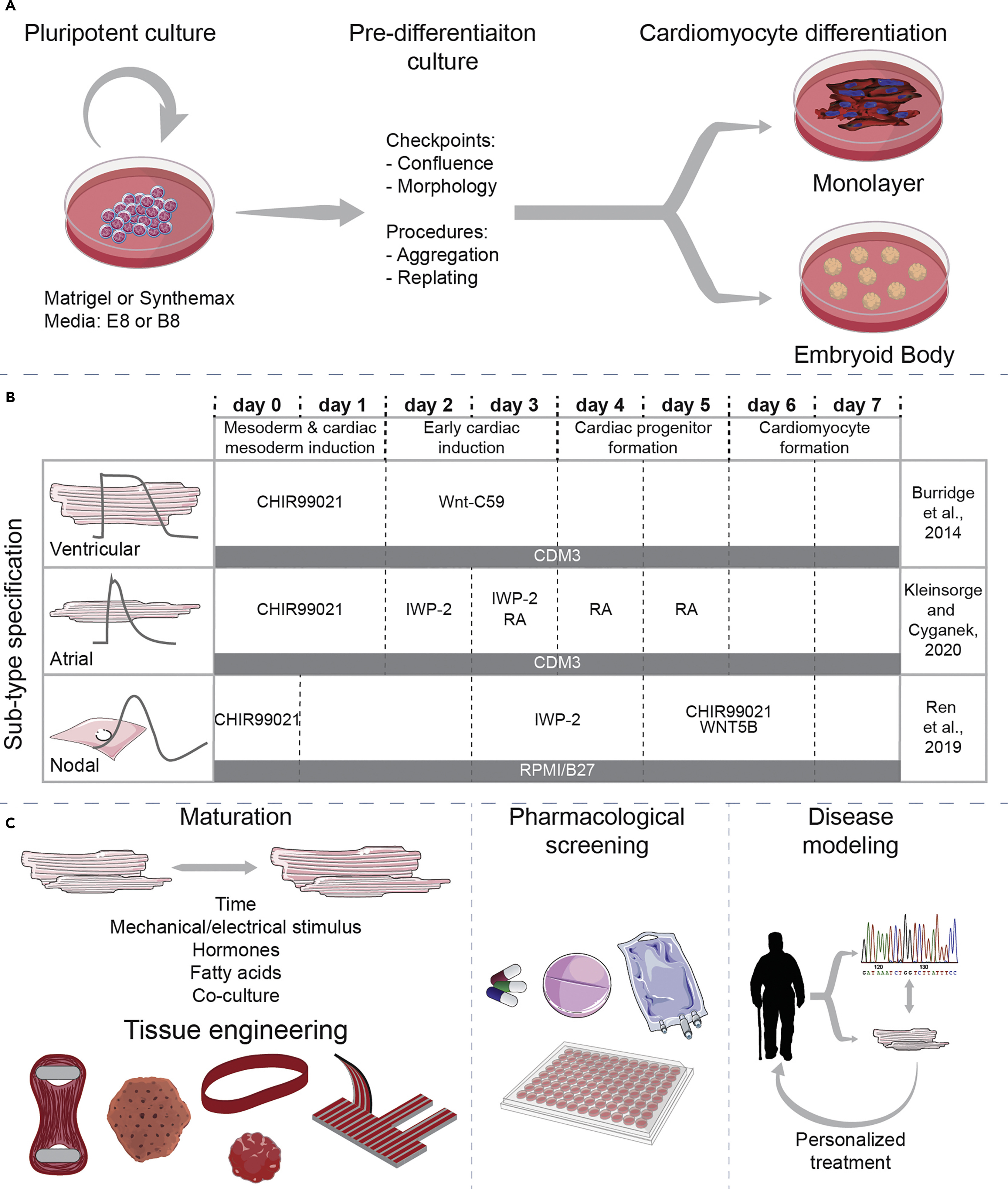
A review of protocols for human iPSC culture, cardiac differentiation, subtype-specification, maturation, and direct reprogramming
The methods for the culture and cardiomyocyte differentiation of human embryonic stem cells, and later human induced pluripotent stem cells (hiPSC), have moved from a complex and uncontrolled systems to simplified and relatively robust protocols, using the knowledge and cues gathered at each step. HiPSC-derived cardiomyocytes have proven to be a useful tool in human disease modelling, drug discovery, developmental biology, and regenerative medicine. In this protocol review, we will highlight the evolution of protocols associated with hPSC culture, cardiomyocyte differentiation, sub-type specification, and cardiomyocyte maturation. We also discuss protocols for somatic cell direct reprogramming to cardiomyocyte-like cells.
In STAR Prot,
2022
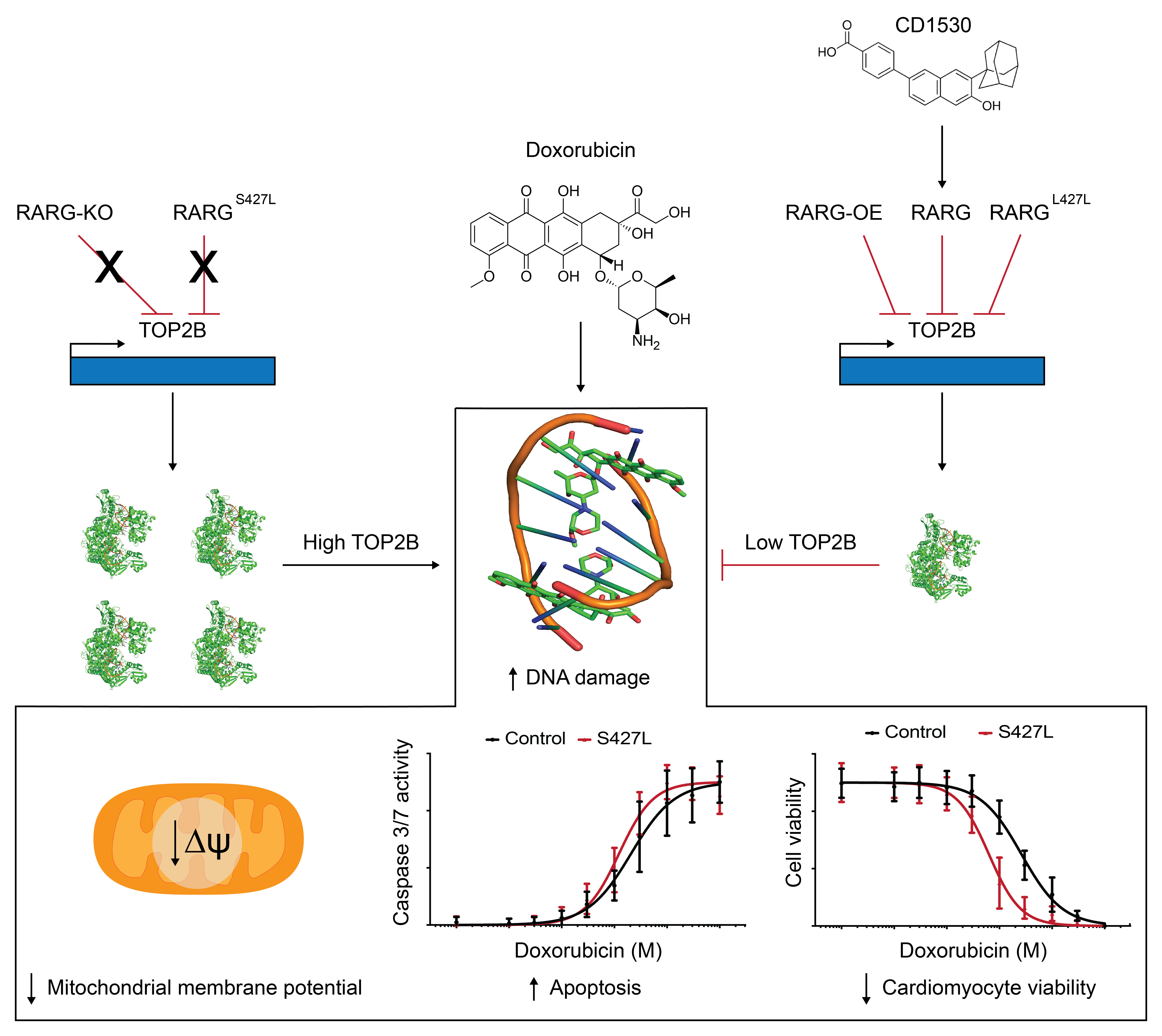
RARG variant predictive of doxorubicin-induced cardiotoxicity identifies a cardioprotective therapy
Doxorubicin is an anthracycline chemotherapy agent effective in treating a wide range of malignancies, but its use is limited by dose-dependent cardiotoxicity. A recent genome-wide association study identified a SNP (rs2229774) in retinoic acid receptor-γ (RARG) as statistically associated with increased risk of anthracycline-induced cardiotoxicity. Here, we show that human induced pluripotent stem cell-derived cardiomyocytes (hiPSC-CMs) from patients with rs2229774 and who suffered doxorubicin-induced cardiotoxicity (DIC) are more sensitive to doxorubicin. We determine that the mechanism of this RARG variant effect is mediated via suppression of topoisomerase 2β (TOP2B) expression and activation of the cardioprotective extracellular regulated kinase (ERK) pathway. We use patient-specific hiPSC-CMs as a drug discovery platform, determining that the RARG agonist CD1530 attenuates DIC, and we confirm this cardioprotective effect in an established in vivo mouse model of DIC. This study provides a rationale for clinical prechemotherapy genetic screening for rs2229774 and a foundation for the clinical use of RARG agonist treatment to protect cancer patients from DIC.
In Cell SC,
2021

Mitochondrial architecture in cardiac myocytes depends on cell shape and matrix rigidity
Contraction of cardiac myocytes depends on energy generated by the mitochondria. During cardiac development and disease, the structure and function of the mitochondrial network in cardiac myocytes is known to remodel in concert with many other factors, including changes in nutrient availability, hemodynamic load, extracellular matrix (ECM) rigidity, cell shape, and maturation of other intracellular structures. However, the independent role of each of these factors on mitochondrial network architecture is poorly understood. In this study, we tested the hypothesis that cell aspect ratio (AR) and ECM rigidity regulate the architecture of the mitochondrial network in cardiac myocytes. To do this, we spin-coated glass coverslips with a soft, moderate, or stiff polymer. Next, we microcontact printed cell-sized rectangles of fibronectin with AR matching cardiac myocytes at various developmental or disease states onto the polymer surface. We then cultured neonatal rat ventricular myocytes on the patterned surfaces and used confocal microscopy and image processing techniques to quantify sarcomeric α-actinin volume, nucleus volume, and mitochondrial volume, surface area, and size distribution. On some substrates, α-actinin volume increased with cell AR but was not affected by ECM rigidity. Nucleus volume was mostly uniform across all conditions. In contrast, mitochondrial volume increased with cell AR on all substrates. Furthermore, mitochondrial surface area to volume ratio decreased as AR increased on all substrates. Large mitochondria were also more prevalent in cardiac myocytes with higher AR. For select AR, mitochondria were also smaller as ECM rigidity increased. Collectively, these results suggest that mitochondrial architecture in cardiac myocytes is strongly influenced by cell shape and moderately influenced by ECM rigidity. These data have important implications for understanding the factors that impact metabolic performance during heart development and disease.
In JMCC,
2020
Mitochondrial Function in Engineered Cardiac Tissues is Regulated by Extracellular Matrix Elasticity and Tissue Alignment
Mitochondria in cardiac myocytes are critical for generating ATP to meet the high metabolic demands associated with sarcomere shortening. Distinct remodeling of mitochondrial structure and function occur in cardiac myocytes in both developmental and pathological settings. However, the factors that underlie these changes are poorly understood. Because remodeling of tissue architecture and extracellular matrix (ECM) elasticity are also hallmarks of ventricular development and disease, we hypothesize that these environmental factors regulate mitochondrial function in cardiac myocytes. To test this, we developed a new procedure to transfer tunable polydimethylsiloxane disks microcontact-printed with fibronectin into cell culture microplates. We cultured Sprague-Dawley neonatal rat ventricular myocytes within the wells, which consistently formed tissues following the printed fibronectin, and measured oxygen consumption rate using a Seahorse extracellular flux analyzer. Our data indicate that parameters associated with baseline metabolism are predominantly regulated by ECM elasticity, whereas the ability of tissues to adapt to metabolic stress is regulated by both ECM elasticity and tissue alignment. Furthermore, bioenergetic health index, which reflects both the positive and negative aspects of oxygen consumption, was highest in aligned tissues on the most rigid substrate, suggesting that overall mitochondrial function is regulated by both ECM elasticity and tissue alignment. Our results demonstrate that mitochondrial function is regulated by both ECM elasticity and myofibril architecture in cardiac myocytes. This provides novel insight into how extracellular cues impact mitochondrial function in the context of cardiac development and disease.
In AJP Heart,
2017